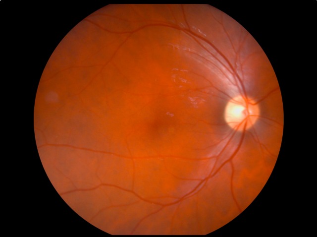
Your retina, a delicate film at the back of your eye, holds the script for nearly every visual memory you cherish. When that script becomes smudged by disease, macular degeneration, diabetic retinopathy, retinitis pigmentosa, the light you see bends and fragments. In the UK alone, diagnoses of sight-threatening retinal diseases are steadily on the rise. You might even know someone whose world has been redrawn in softer edges because of these conditions.
The burden reaches well beyond the personal: healthcare, social services, and the working world feel the strain. Retinal diseases can erase independence: they twist the day-to-day for patients and families. Yet, progress flickers at the frontier. To push back against this shadow, research needs a way to emulate the real thing. That quest for authenticity, mirroring disease in a controlled way, defines the conversation around retinal disease modelling.
Traditional Models of Retinal Disease
Picture this: before the age of organoids and stem cells, your main tools for probing retinal disease looked very different. Researchers leant heavily on histological techniques, glass slides under a microscope, preserved retinas showcasing fixed pathology. You often find that these approaches, while valuable, freeze the story mid-chapter.
In some cases, animal eyes from species with similar retinal structure, like rabbits or pigs, offered crucial clues, though with caveats. You might see artificial lesions created with lasers or chemicals: these mimic parts of disease but rarely capture the full drama. Clinical observation in humans, laborious record-keeping, and drawing comparisons between healthy and impaired vision have played their roles as well. Yet, statistical models, while helpful, can feel abstract, missing the nuance and unpredictability of living tissue.
In Vitro Models for Retinal Disease
You will find that modern in vitro models have reimagined the laboratory bench, swapping static slides for living systems. Cell cultures, whether derived from retinal pigment epithelium or differentiated from induced pluripotent stem cells, offer you a microscopic stage to replay disease events. These methods might involve exposing cells to stressors that echo the challenges of diabetic environments or oxygen starvation.
Three-dimensional organoids, sometimes described as mini-retinas, provide an impressive lens into development and degeneration. These clusters of human cells grown in dishes move beyond flatness, developing layers similar to your own retina. With them, you can observe how specific genes or toxins ignite the spiral of cell loss. You will see interactions between various retinal cell types play out in real time under different experimental cues.
While in vitro models grant unmatched control and a human genetic context, they can lack the whole-organism subtleties: blood flow, immune response, and neural connections beyond the eye remain out of focus.
In Vivo Animal Models and Their Contributions
When your question turns to whole-body responses and progression over time, animal models often step into the spotlight. Mice dominate the scene thanks to specific gene editing tools, knockouts, knock-ins, allowing you to create precise versions of human conditions. Zebrafish, with their transparent embryos and rapid development, bring surprising clarity to early disease stages: you can watch retinal cells decline or recover with each passing hour.
Larger mammals sometimes stand in for the human eye’s architecture, mimicking blood vessels or cell types more faithfully. Through these models, treatments like gene therapy and novel pharmaceuticals take their first steps towards human trials. You will find they can even help unpick the chain of inflammatory responses or track the migration of stem cells transplanted into a damaged retina.
Nevertheless, animal models may mislead as often as they enlighten: species differences and ethical dilemmas always cloud the findings. Predicting human response by starting with a mouse or fish can turn progress into guesswork.
Emerging Approaches in Retinal Disease Modelling
A new wave of modelling has arrived, born of your era’s appetite for precision and complexity. Bioengineered retinas, crafted from patient-specific stem cells, open a door onto personalised medicine. Here, every chip of tissue is shaped by the defects and history unique to one person, tightening the lens on prediction.
Computational models and machine learning, once outsiders, now stand shoulder to shoulder with biologists. You might not expect numbers to simulate sight loss, but algorithmic models can map molecular pathways and even forecast how patients respond to new interventions. Virtual retinas on a computer screen blend real-world data with simulation, offering a glimpse of trials that haven’t happened yet.
Meanwhile, organ-on-chip technologies weave blood vessels and nerve inputs into miniaturised living systems, capturing the push and pull between different eye structures. These hybrid methods promise to unite the detail of in vitro with the breadth of in vivo, giving you nuanced pictures where black-and-white once prevailed.
Challenges and Limitations in Current Retinal Disease Models
Even the finest models, for all their ingenuity, keep you guessing. No laboratory retina yet mirrors the unpredictable, sometimes chaotic dance of disease in humans. Genetic background, age, environment, all twist the script. You might spot a therapy that works wonders on cells in a dish, only for it to stumble when faced with the rhythms of a living organism.
Models often struggle to reflect chronic degeneration, slow inflammation or the contribution of the brain to visual changes. Ethical shadows loom large as well: animal models are closely scrutinised and human-derived tissues confront regulatory complexities. Besides, scaling up remains tricky. What thrives in a single experiment sometimes resists mass production or clinical translation.
These constraints remind you that, for all progress, humility still has a place in vision research. The unknown outpaces the known more often than anyone wants to admit.
Future Directions for Retinal Disease Modelling
You will find that hope is stitched through the future of retinal disease modelling. Integrating multiple model types, melding computational predictions with patient-derived organoids, looks set to sharpen accuracy. New imaging techniques promise to capture changes at the sub-cellular level as disease unfolds, catching patterns that eluded even the sharpest eye before.
Gene editing tools, like CRISPR, might enable you to tweak models in a way that mimics rare human mutations with uncanny fidelity. Collaborations across disciplines, data science, pharmacology, ophthalmology, will become standard fare, encouraging creative leaps. One can envision a world where modelling not only tests treatments but predicts disease before a single symptom emerges.
Ethical innovation, too, will march forward. Researchers hope to devise methods that reduce reliance on animals while enhancing relevance to patients. If you keep your ear to the ground, whispers of new discoveries come quick, and surprises, delightful or sobering, will remain inevitable.
To Wrap Up
You have wandered through a shifting landscape, constantly evolving yet full of unanswered questions. Retinal disease modelling, for all its achievements and frustrations, continues to shape what sight might mean in years ahead. Pause for a second, whose vision will the next model save? Maybe yours, maybe someone you’ll never meet. Either way, in the case that you decide to look a little closer at the way science sketches out solutions, you will find that fresh perspectives are always flickering at the edge of light.